
Clinical Education & Research
A centralized resource for guidelines, injury grading, and the global BTAI registry.
Traumatic Aortic Injury (TAI)
Traumatic Aortic Injury remains the second most common cause of death in patients suffering from blunt trauma, second only to head injury. While traditionally associated with high-speed motor vehicle accidents, the changing demographics of the population have seen a rise in injuries related to falls in the elderly.
Epidemiology
- •Incidence: Occurs in approximately 1.5% to 2% of patients with severe blunt thoracic trauma.
- •Pre-hospital Mortality: Historically reported as high as 80% at the scene, though improved EMS transport and resuscitation have increased hospital arrival rates.
Management Trends
- •Paradigm Shift: Over the last two decades, management has shifted from open surgical repair to Thoracic Endovascular Aortic Repair (TEVAR).
- •Medical Management: Increasing evidence supports non-operative management for Grade I (intimal tear) and Grade II (intramural hematoma) injuries.
Injury Subtypes & Clinical Considerations
Blunt Thoracic Aortic Injury (BTAI)
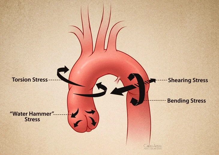
The most common form, typically caused by rapid deceleration (MVCs, falls). The aortic isthmus is the site of injury in >85% of cases due to ligamentum arteriosum tethering.
Key Focus: TEVAR ConformabilityPenetrating Aortic Injury
Caused by GSW or stab wounds. Unlike BTAI, these injuries lack adventitial containment and frequently present with massive hemothorax and shock.
Key Focus: Immediate ControlPediatric TAI
Rare but challenging due to small vessel diameters and future growth. TEVAR carries long-term risks of aortic coarctation as the child grows.
Key Focus: Conservative vs. RepairGeriatric TAI
Elderly patients often present with "shaggy aortas" or severe calcification. The risk of stroke during wire manipulation is significantly higher.
Key Focus: Stroke PreventionMechanism of Injury
The classic mechanism involves rapid deceleration — a frontal motor vehicle collision or fall from height.
Society for Vascular Surgery Injury Grading System
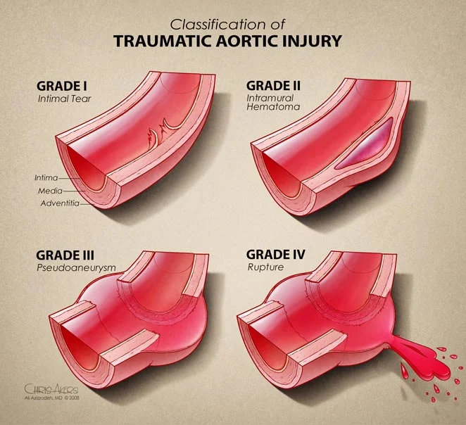
Disruption of the innermost layer only. No external contour abnormality.
Blood collection within the media. May show subtle contour changes but no pseudoaneurysm.
Disruption of intima and media, contained by adventitia. Clear bulge on CTA.
Full-thickness disruption with active contrast extravasation into the chest.
Management Guidelines
Reflects The Society for Vascular Surgery clinical practice guideline on the management of blunt thoracic aortic injury: Focused update (March 2026) — 13 recommendations, the first update since 2011. The guideline writing group was chaired by ATF Chairman Ali Azizzadeh, MD, with ATF President Joseph J. DuBose, MD as lead author.Read the guideline (JVS) ↗
| Injury Grade | Treatment Strategy | Timing & Follow-up |
|---|---|---|
| Grade 1 | Nonoperative management | Definitive medical therapy. No routine follow-up imaging. |
| Grade 2 | Nonoperative management | Definitive medical therapy. At least one follow-up CTA at 1–3 months to confirm resolution. |
| Grade 3 | TEVAR | Stable: delayed TEVAR (>24 h) suggested, to allow management of associated injuries. Unstable (BTAI the specific cause of instability): urgent (<24 h) or emergent. |
| Grade 4 | TEVAR | Emergent repair |
// Grades 1 and 2 together constitute minimal aortic injury (MAI).
Imaging Surveillance
- •Grade 1 (NOM): routine surveillance imaging is suggested against.
- •Grade 2 (NOM): at least one follow-up study is suggested, with repeat imaging at 1–3 months after injury.
- •Post-TEVAR: postoperative surveillance imaging is suggested. The interval is individualized rather than fixed.
Concomitant TBI or Solid Organ Injury
- •Grade 1–2 with TBI: anti-impulse therapy for the BTAI is suggested against. Blood pressure management should prioritize the TBI.
- •Grade 3 with TBI: anti-impulse therapy individualized through collaborative management with other specialties.
- •Grade 3 with TBI or SOI: TEVAR timing determined collaboratively, in consultation with trauma and neurosurgery.
- •Grade 4: emergent repair is recommended.
Left Subclavian Artery Coverage
Among patients requiring LSA coverage (zone 2) for TEVAR, the decision on revascularization is suggested to be based on feasibility and on factors such as a patent prior left internal mammary–to–coronary bypass, a dominant left vertebral artery on pre- or intraoperative imaging, or an aortic origin of the left vertebral artery.
Intraoperative Anticoagulation
Intraoperative anticoagulation is suggested to be used at the surgeon's discretion, weighing the risk of bleeding against thrombotic complications.
Where TBI or SOI coexists, a collaborative approach is suggested. Observational data offer some reassurance that heparinization is not associated with worse intracranial bleeding or neurologic outcomes, but the certainty of that evidence is very low.
Anti-impulse Therapy in Nonoperative Management
In Grade 3 BTAI, anti-impulse therapy is suggested as a stabilizing measure until TEVAR is performed, provided concomitant injuries do not preclude it. In Grade 1 and 2 injuries with concomitant TBI, it is suggested against.
Knowledge Gaps & Future Directions
Despite these guidelines, significant questions remain regarding the long-term durability of stent grafts in young trauma patients, the optimal management of Grade II injuries, and the impact of left subclavian artery coverage.
The Aortic Trauma Foundation BTAI Registry was established to answer these critical questions through multi-center collaboration.
Clinical disclaimer
This material is provided for professional education and reference. It is not a substitute for clinical judgment, institutional protocol, or the current full text of the applicable practice guidelines. Clinicians should consult the primary literature and the most recent Society for Vascular Surgery guidance when making management decisions.